ORL
This area of expertise of the otolaryngology specialist focuses on the ear-central nervous system interface. The ear, the central nervous system and their related structures function synchronously in the production and processing of information that we interpret as sound, balance or posture. Oto-neurology concerns the diagnosis and treatment of a variety of pathologies: hypoacusis, tinnitus, vestibular disorders, inflammatory diseases, malformations or auricular tumors, lesions of the facial and acoustic-vestibular nerves, diseases of the skull base.
ORL Investigations
-
Computerized posturography
Computerized posturography is a computerized clinical trial, which takes 45 minutes, and is used for balance assessment. The components used to maintain balance are: the muscles, the proprioceptive system, the portion of the vestibular system and the vision. Posturographic tests isolate each of these components to assess the location of the deficit, if any. The test also assesses the ability of the autonomous motor system to recover from sudden and unexpected movements.
During the test, the patient is positioned in the center of a platform, facing a screen on which various images are projected. In many cases, the platform or wall is manipulated depending on the patient's movements. Patient data are entered into the computer before testing, in order to compare the vestibular, visual and proprioceptive system responses with those corresponding to age.
Posturography is also a very useful investigation to determine a baseline for vestibular rehabilitation therapy and in designing an individual treatment program. Once recovery therapy is initiated, posturography can be used to evaluate patient progress by comparing post-therapy testing with baseline assessment.
- Diagnostic tool for balance disorders
- Highly suggestive for the cause of balance disorder
- Proprioceptive deficit: Spine pathology, muscle diseases, B12 deficiency
- Visual deficit (patients who rely almost exclusively on visual analyzer to maintain posture)
- Deficiency of the vestibular apparatus (vertigo, peripheral and central vestibular disorders)
- Highlights the central integration deficit of the 3 analyzers
- Evaluates the risk of falling in patients with balance disorders, regardless of the cause (except lipotimes, syncopes, which are not balance disorders)
- Detects simulators - the response to changing posture is reflex, it cannot be simulated
- Treatment: posturographically assisted vestibular rehabilitation
- Peripheral vestibular syndromes with balance disorders outside the vestibular crisis (Meniere's disease, post vestibular neuronitis, destructive vestibular syndromes of vascular origin)
- Central vestibular syndromes
- After trauma with damage to the temporal rock
- After surgery with lesion/destruction of the labyrinth, vestibular nerve
- Diseases of the osteo-articular system, diabetes mellitus with sensitivity disorders
- The patient learns to compensate for the deficit.
-
VEMP
Vestibular myogenic evoked potentials (VEMP) Determining vestibular myogenic evoked potentials is an exploration used to evaluate the function of the structures of the internal ear involved in maintaining balance (utricula, sacula, upper vestibular nerve, inferior vestibular nerve and their central connections). VEMPs can be determined by placing electrodes on different muscles during contraction, hence the name of cervical VEMP (sternocleidomastoid muscle) - cVEMP and ocular VEMP (lower oblique muscle) - oVEMP. cVEMP evaluates the function of the sacula and lower vestibular nerve. The sacula, the lower portion of the motion sensor in the inner ear, is sensitive to vertical (top to bottom) movements of the head and in response to these movements sends impulses along the lower vestibular nerve. These impulses control the movements of the head by relaxing the sternocleidomastoid muscles (SCM).
VEMP testing involves attaching electrodes to the patient's forehead, cheeks or neck, which will be positioned horizontally. Also, in each ear a helmet will be placed, through which a sound stimulus is transmitted, followed by the electrodes recording the response of the muscle to the vestibular stimulus.
-
PEA
Testing of the auditory evoked potentials is used to evaluate the hearing and to identify abnormalities of the auditory nerve and auditory pathways up to the brainstem.
Recommendations:
- Asymmetric or unilateral deafness: screening for acoustic neurinoma, diagnostic information in demyelinating diseases (multiple sclerosis)
- Provides qualitative and quantitative information regarding the function of the inner ear and the auditory nerve
During the test, the patient is positioned horizontally. Electrodes are placed on the forehead and behind each ear and in the earphones for the sound stimulus, perceived as a brief click. The electrodes perceive the brain's responses to these sounds and record them. Patients should not respond to the click, but are invited to remain as calm and relaxed as possible.
Abnormal results may be a sign of hearing loss, multiple sclerosis, acoustic nerve neuroma, stroke. Abnormal outcomes may also be due to: brain trauma, brain malformations or brain tumors. -
Video Frenzel
This examination is used for the evaluation of nystagmus with the help of special glasses fitted with an infrared camera. These glasses are connected to a video monitor that the investigator watches the eye movements and the appearance of nystagmus.
- In the absence of fixation (in the dark) - the nystagmus characteristic of peripheral vestibular syndromes is detected
- At fixation: detecting central vestibular syndromes
- Characterization of nystagmus: direction, duration, latency
- Nystagmus occurring during provocative maneuvers: diagnosis of benign positional vertigo
- Spontaneous nystagmus - the patient is asked to look in different directions without moving his head
- Nystagmus revealed
- Head thrust test: During the test the Frenzel eyeglass cover is removed and the patient is asked to look to the right or left by turning the head or fixing an object. The investigator will suddenly turn his head in the opposite direction, following the appearance of nystagmus;
- Head-shaking test: the investigator mobilizes the patient's head horizontally, vertically or frontally for 20 seconds, following which the appearance of nystagmus is followed;
- Dix-Hallpike maneuver: the patient is brought from the sitting position in the dorsal decubitus with the head rotated at 45 degrees and in extension at about 20 degrees. The test is positive if nystagmus appears.
-
ASSR
Auditory Steady State Response is a new test that is being conducted with ABR to assess hearing.
Recommendations: infants, young children, people unable to respond to the conventional audiogram.
Medical-legal instrument: it is an objective measurement - the detection of simulants, the objective evaluation of hearing in people working in a noisy environment
The tested person should be relaxed and stay as still as possible to get reliable results. Most of the time, the tests are performed under sedation or during natural sleep, if the person is under 6 months. The results are obtained by measuring the brain activity while the person listens to tones of different frequencies and intensities. The brain activity is recorded by means of electrodes attached to the forehead and behind each ear. The use of electrodes eliminates the need for active patient participation (for example, pressing a button in response each time a tone is heard). The results are obtained objectively using statistical formulas that determine the presence or absence of a real answer.
-
Acoustic ottoemissions
Acoustic ottoemissions are low-pitched sounds produced by the outer ciliary cells of the inner ear (cochlea). They may occur spontaneously or in response to clicks or tones. When the cochlear cells in the cochlea are stimulated, they respond by sending information to the brain and sending an "echo" back to the outer ear. This "echo" can be analyzed and recorded. OAEs are usually present in people with normal hearing function, but may be absent if there is even mild transmission or sensory hearing loss.
OAE testing is useful in:
- screening for hearing loss in newborns, young infants;
- Identification of functional or inorganic hearing loss (simulants);
- differential diagnosis between cochlear and retrocochlear sudity;
- monitoring the effect of drugs with toxic potential, being able to detect the cochlear dysfunction before hearing loss;
- may provide objective confirmation of cochlear dysfunction in patients with normal hearing;
- it may be an early warning sign of cochlear dysfunction induced by noise exposure;
- may provide information on cochlear function in patients with tinnitus by identifying the cochlear region corresponding to tinnitus frequency.
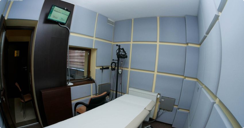
Testing is achieved in a soundproofed room. During testing, a soft rubber or silicone probe is sealed in the ear canal. A series of clicks or tones are delivered through the probe tip. At the same time the probe perceives the very fine sound that represents the "echo" or the response of the cochlea. It is important for the patient to remain still and to remain relaxed and calm during the test. The records are then printed and analyzed. The test duration is about 10 minutes.
-
Electrocohleography
Electrocochleography is indicated in the diagnosis of Meniere's disease. This test is an objective way of measuring the electrical potentials generated in the inner ear as a result of sound stimulation. This test is most often used to determine if there is an increase in pressure of the endolymph in the cochlea. Increased fluid pressure in the cochlea may cause symptoms such as hearing loss, sensation of fullness in the ear, dizziness and/or tinnitus. These symptoms are the expression of diseases of the inner ear, such as Meniere's disease or endolymphatic hydrops.
A patient undergoing an electrocochleography test will have multiple electrodes placed on the scalp. A small electrode and a foam headset will then be placed in the external ear canal of the tested ear. The patient will be instructed to relax, listening to a clicking sound. It is very important for the patient to be relaxed for this test, as any movement can disrupt the evaluation. The patient does not have to respond to the sound stimulus. The electrocochleography lasts up to 40 minutes.
-
Audiogram
Tonal and vocal audiometry
- Diagnosis of neuro-sensory and transmission hearing loss
- Periodic evaluation of people at risk of deafness: diabetes, autoimmune diseases, vasculitis
- When appropriate, hearing aid recommendation
High Frequency Audiometry:
- highlighting hearing loss for frequencies over 8 kHz in patients with tinnitus and normal hearing on conventional audiograms
- TEN test - highlighting areas of the cochlea that have no hearing function (useful for calibrating the hearing aid)
During the hearing test, the patient will be placed a pair of headphones on the ears, along with a band that supports a bone conductor on the scalp, as the threshold for tones is measured on a frequency range. Typically, the range is 250 - 8000 Hz, as it encompasses the range of speech frequencies. The voice audiogram includes two tests. First, speech recognition threshold (SRT) is used to measure the lowest level at which words can be repeated. Usually words with two syllables are used, with equal emphasis on each word. The second test, the speech discrimination test, is used to evaluate the ability to understand and repeat words with a single syllable presented at a high volume.
The high frequency audiogram evaluates the threshold for tones above 8000 Hz (8-20 kHz). It is indicated for people exposed to noise and people with tinnitus.
TEN ("threshold equalizing noise") is a new test used to determine non-functional regions of the inner ear.
-
Endoscopy
It is defined as the visual examination of a cavity organ using the endoscope. The endoscope is a thin tube (rigid or flexible) equipped with a series of lenses and illuminated by fiber optics. The outer end of this tube can be attached to a camcorder connected to a monitor. Thus, the examined area is viewed on the monitor.
Recommendations:
- Confirmation/rejection of the presence of rhinosinusitis by highlighting muco-purulent secretion in the middle meatus.
- Diagnosis of nasal polyposis
- Confirmation/rejection of the presence of obstructive adenoid vegetation
- Confirmation/rejection of rhinopharyngeal tumor pathology (especially young people with chronic unilateral otitis)
- Diagnosis of functional dysphonia, laryngitis, vocal nodules, gastroesophageal reflux disease with pharyngeal laryngeal reflux, rhinopharyngeal tumor pathology, vocal cord paralysis
Oto-endoscopy is an extremely useful exploration in the outpatient evaluation of the ear. The only requirement that the patient must meet during the examination is to remain still. The examination is not painful and does not require prior local anesthesia. The endoscope offers a wide field of view, the eardrum can be examined entirely and its mobility can also be evaluated. The presence of fluid in the middle ear can be visualized much better with the endoscope than with the otoscope, and the integrity of the middle ear tufts can be assessed through the perforated eardrum. Otto-endoscopy offers a better visualization of retraction bags compared to conventional otoscopy, offers a better evaluation of the operated ear and post-mastoidectomy cavities (operation for cholesteatoma). It is also an extremely useful tool in performing outpatient procedures such as: auricular toilet, extraction of the impacted cerumen plug, aspiration of ear secretions (otitis), removal of foreign auricular bodies.
Nasal endoscopy offers a detailed examination of the nostrils and the rhinopharynx. The most common reasons for this examination are nasal obstruction, suspected acute or chronic sinusitis, nasal/facial pain, odor disorders, and epistaxis. This test allows a full and detailed view of nasal mucous, nasal cornets, communication with para-nasal sinuses and rhyno-pharinx.
Rigid laryngeal endoscopy
It allows for a detailed view of the larynx and the lower portion of the pharynx. The procedure is performed with the patient in a sitting position. The lens of the endoscope, oriented lower oblique, will allow the visualization of the larynx and the hypopharynx.Flexible fibroscopy
It allows the evaluation of nostrils, pharynx and larynx with a small flexible tube. It can be used for examining nostrils, but is most often used for visualizing the larynx.


